
the brain, cell by cell
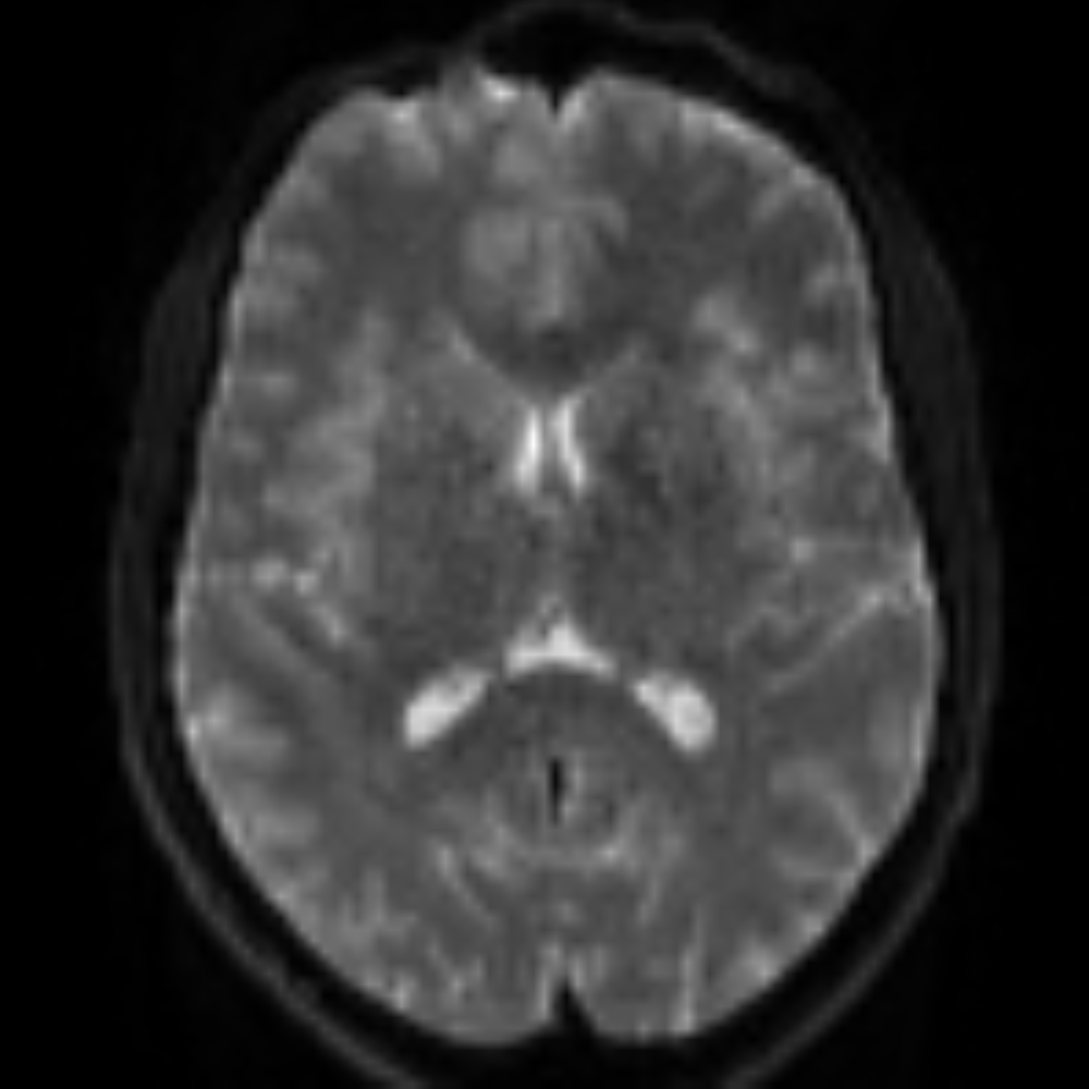
MRmicro derives quantitative maps of axonal density, neurite geometry and gray-matter cytoarchitecture — biomarkers sensitive to MS, dementia and traumatic injury, more specific than conventional MRI.
MRmicro turns clinical diffusion MRI into quantitative brain microstructure maps, supporting assessment of axonal integrity, cortical tissue composition and disease-related change in neurological workflows.
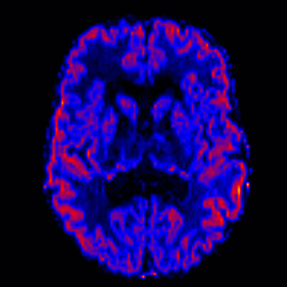
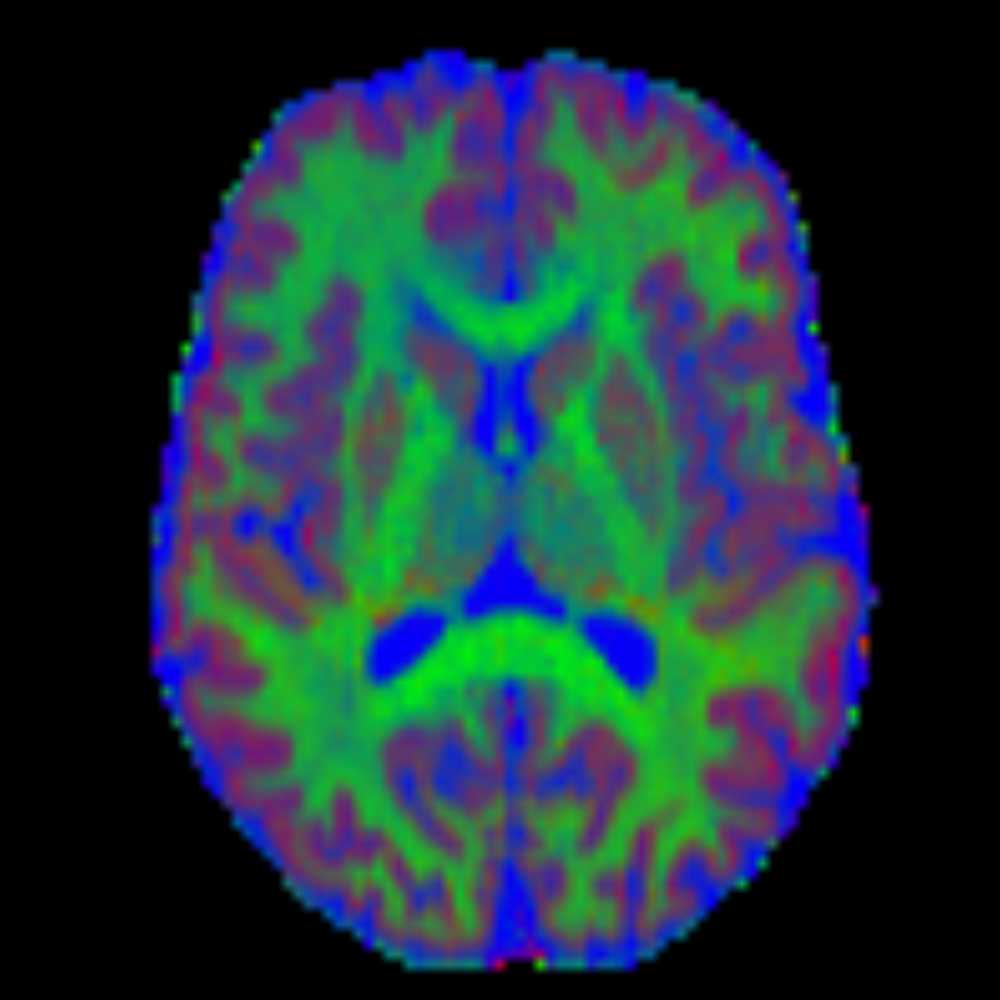
cell-body fraction
Cortical microstructure at millimetre scale, designed to read cell-body density on clinical 3T acquisitions. Sensitive to early neurodegeneration and cortical dysplasia.
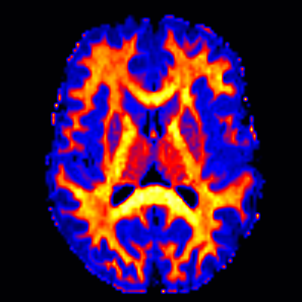
intra-neurite fraction
Voxel-wise integrity of axons and dendrites across white matter. Picks up demyelination in tissue that still looks normal on FLAIR and tracks EDSS progression in MS.
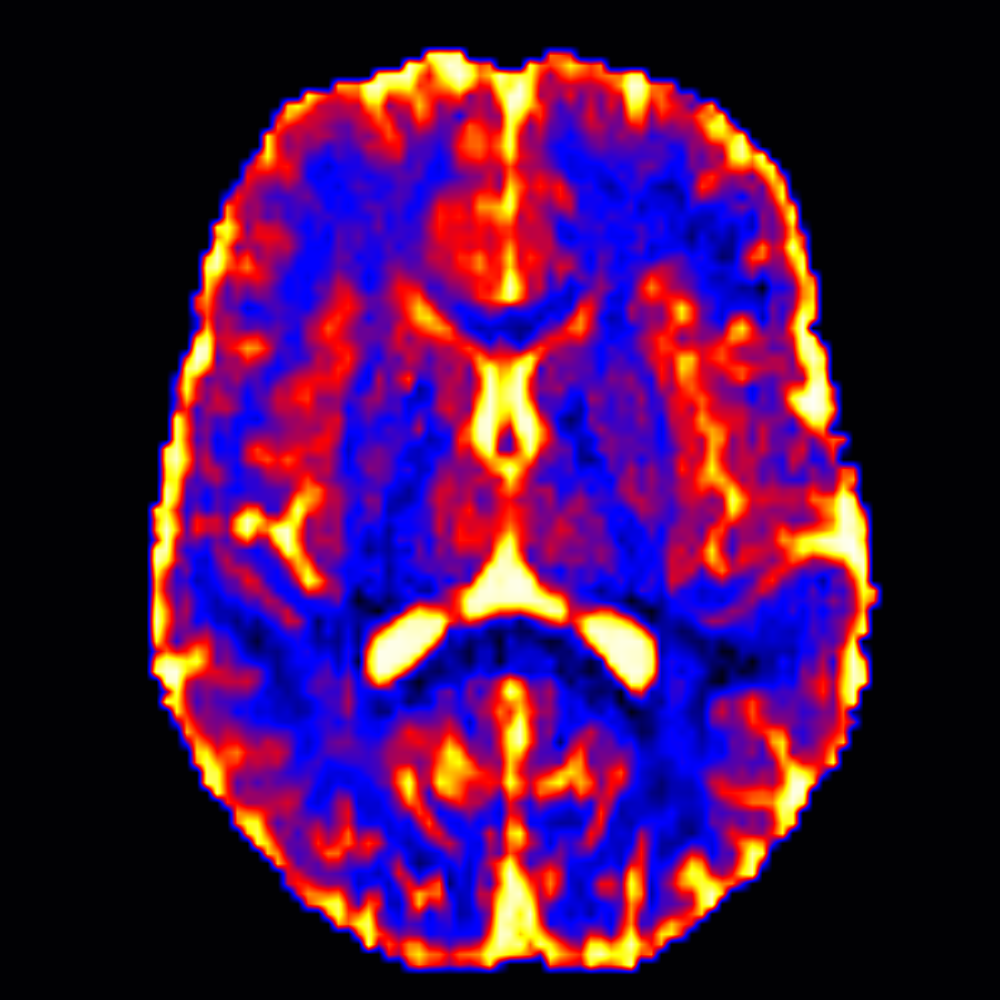
extra-cellular fraction
Free water and interstitial space between cells. Rises with edema, neuroinflammation and tissue rarefaction, making it an early marker of neurodegeneration.
Lightweight layer Between your scanner and your PACS.
MRmicro runs on a standard workstation. No cloud. No data leaves the hospital.
Scanner
Any 1.5 T or 3 T system from any vendor. Based on multi-shell DWI sequence.
DICOM router
Hospital network routes diffusion series to MRmicro.
MRmicro node
Local workstation. Full processing in 3 minutes.
Derived series
Parametric maps written as standard DICOM, sent back to PACS.
Reading room
Radiologist opens the maps in the existing viewer. No new software to learn.